
Signed in as:
filler@godaddy.com

Poster 1
Poster 2
Poster 3
Poster 4
Poster 5
Poster 7
Poster 8
Poster 9
Poster 10
Poster 11
Poster 12
Poster 13
Poster 14
Poster 6
Viktoriia Yerokhina
School of Medicine, University of Central Lancashire,
Burnley, United Kingdom
X: @vi_histo
I was always good at biology, which is why I decided to study Medicine in Ukraine. I was a diligent student who always completed homework, and my peers often asked me to explain the content. I never rejected the opportunity to help struggling students because even then, without knowing pedagogy or Dale's ‘Cone of Learning,’ I felt that teaching was the best way to learn. Throughout my education, my lecturers consistently expressed that I possess natural teaching skills and encouraged me to consider returning for a teaching role in the future. Eventually, I realised that I not only enjoy teaching but also find immense satisfaction in witnessing the 'aha' moment in someone's eyes when they grasp a concept. After graduating from university in Ukraine in 2012, I began lecturing in histology, cytology, and embryology to international students, sharing best practices with colleagues initially in Ukraine and later in the UK. In 2015, I completed my PhD in anatomy and continued my path in academia. After ten successful years of teaching, in September 2022, I was forced to move from Ukraine to the UK to teach medical sciences due to the ongoing war. My positive mindset accepts every challenge as an opportunity, which is why I continue my academic journey. In 2023, I won the nomination for Best Lecturer at the University of Central Lancashire and was shortlisted for the award again in 2024. My passion for helping learners and fostering their understanding has become a central aspect of my life.
Keywords: academic journey, career reflection, anatomy lecturer
Alice Brown, Naila Ali
Medical and Dental Science, University of Birmingham,
Birmingham, United Kingdom.
Hypertension and its downstream complications often arise due to the lack of awareness and education surrounding risk factors and consequences. Previous studies have shown the efficacy of Three-Dimensional models in disease education among patients and medical students, however, evidence of its use within the general public is limited. Hence, a two-armed unblinded randomised control trial was undertaken to investigate 3D models in educating non-science university students (n=20) about HTN and Atherosclerosis. Following randomisation, participants underwent an educational intervention using either Two-Dimensional Images (n=9) or 3D models (n=11). Using methods of pre- and post-intervention assessments, a Likert scale and a free text comment section; the differences in knowledge, confidence, self-efficacy and individual experience between the two groups were evaluated. The findings indicate that 3D models are comparable to 2D images, though higher knowledge scores were reported there was no statistical difference (p=0.22). Subjective content confidence and self-efficacy were high in both groups but no differences between arms (p>0.05), moreover, the 3D models showed significantly higher engagement and understanding (p<0.05). Quantitative and qualitative analyses provide evidence of the high visualisation, spatial navigation and enjoyment implored by 3D models. This pilot RCT provides evidence for 3D model implementation in disease awareness but identifies the need for further research into a larger more representative sample population and differences in long-term knowledge retention, attitude and behavioural changes.
Keywords: 3D models, hypertension, andragogy
Ethics statement: Approved by the University of Birmingham School of Biomedical Science Ethics Committee (Project No: BMSRP_2024_005).
Ethan Jones-Patel
University of Sunderland, School of Medicine,
Sunderland, United Kingdom
bi53gg@student.sunderland.ac.uk
Recurrent Laryngeal nerve Injury (RLNI) is a notable complication of thyroid surgery due to its complex anatomical position and variation that can require further medical intervention. The Recurrent Laryngeal Nerve (RLN), crucial for laryngeal function, follows a convoluted path in the neck with significant anatomical variations across individuals and demographics. Surgical procedures can damage the RLN, resulting in complications such as vocal cord paralysis. The aims of this project are to review the literature from The National Library of Medicine and ResearchGate from 2014 to 2024 to understand the anatomical variation of RLN and the epidemiology of RLNI along with evidence-based measures. Epidemiological factors involving patients of black ethnicity aged 65 years old or over are at greater risk of RLNI. However, differences in gender and Body Mass Index (BMI) are not at greater risk of RLNI. Epidemiological aspects and type of thyroid surgery did not affect the mechanism of injury (MOI), with traction and thermal injuries being the most common. Variation of the RLN is common, with six variations were identified. Identifying the RLN and its variations is often difficult peri-operatively due to inflammation. Prevention of RLNI is critical in avoiding transient or permanent Vocal Cord Palsy (VCP). Injury prevention currently relies on uses Intra-Operative Nerve Monitoring (IONM) and visualisation techniques, with future directions involving rigorous surgical training and minimally invasive robotic surgery. IONM and VA have not been proven to be more effective than each other, but both reduce the rate of VCP.
Keywords: recurrent laryngeal nerve, thyroid surgery, vocal cord palsy
Monisha Tarini Premkumar, Samantha Goodchild
Anglia Ruskin University, School of Medicine,
Chelmsford, United Kingdom
Cadaveric dissection is a centuries-old human anatomy learning resource. Traditionally, formaldehyde caused student-wellbeing concerns. More recently, the WhitWell embalming method has been pedagogic of colour, smell, texture, joint mobility and reduced costs, while improving student experience of haptic and visual tissue characteristics. Our aim is to explore the impact of WhitWell (1.5% HCHO) embalmment in axillary cadaveric dissection on medical students’ performance, experience and satisfaction. Dissected axilla region of a WhitWell preserved cadaver will be used for structure identification using a checklist. Likert Scale and thematic analysis will be used to analyse experience and satisfaction. Ethical approval is ongoing. Williams et al (2018) reported that students’ maximum mean axillary dissection difficulty was a score of 7.5/10 (SD=2.3) and this had practical examination performance implications. The axillary region is a clinically significant area to learn due to the fact that 55920 new cases of breast cancer in the UK are reported between 2016-2018 with an 18% increase incidence of breast cancer since 1983. Therefore, mastectomy and lymph node dissections in breast cancer are lifesaving managements with an injury risk to long thoracic nerve causing winged scapula. Students’ knowing axillary region anatomy with clarity and confidence will help to save lives and reduce surgical complications. Another common procedure done by physicians is a chest drain insertion which necessitates knowing the axillary anatomy without doubts. WhitWell could vastly enhance student anatomy understanding, this research is a step forward in that direction.
Keywords: WhitWell’, anatomy, cadaveric dissection
Ethics statement: Ethics number: ETH2324-6879.
Benedicta Quaye
Department Of Clinical Anatomy, Faculty of Health and Medicine, Lancaster University,
Lancaster, United Kingdom
My foremost encounter with an anatomist was with a surgeon who taught human anatomy during my undergraduate degree. He used no PowerPoint or slides during lectures, and I was always fascinated how much anatomy he effortlessly delivered during every interaction. This ignited my interest further in the discipline. After my first degree, I accepted my first teaching role in anatomy and physiology to Nursing students and realised the more I taught the more I enjoyed the discipline. Indeed, Anatomy is learning about your own body and only as complicated as you make it. I decided to build my career and make a profession out of this discipline. Formally, my career as an anatomist begun after I enrolled into an MPhil in human Anatomy at the University of Ghana Medical School. After my postgraduate degree in Anatomy, my love for anatomy kept growing trough participation in both practical and theoretical teaching of various students focusing on the 3 main branches of the discipline (Embryology, Histology and Gross). After a while, I decided to go further and pursue a terminal degree, and this took me to Germany where I was able to successful complete two PhDs in anatomy and experience teaching anatomy the Deutsch way. In a permanent position now and the journey so far has been an enlightening and maturing one. The ever-changing face of anatomy motivates me to evolve with the times, so I can be part of the gate keepers ensuring anatomy stays relevant in medical education.
Keywords: anatomy, career, reflections
Andrew Marken
St George’s University of London,
London, United Kingdom
X: @drewmarken
Andrew could not attend the conference due to unforeseen circumstances, so the poster was presented in his absence.
Photogrammetry is the technique of using multiple photographs of an object to produce a highly spatially accurate 3D model. 3D models are increasingly used in anatomical sciences for teaching and research purposes. In comparison to other methods of creating 3D models from anatomical specimens, photogrammetry does not require specialist equipment and as such offers a low barrier of entry for use. As such, this makes photogrammetry of particular use to early career anatomists, student projects, and institutions with a limited budget who wish to experiment with 3D image capturing. Whilst the creation of 3D models using photogrammetry is in theory accessible, in practice this is often not the case. Limited guides on how to create 3D anatomical specimens using photogrammetry exist and there is often the assumption that researchers will have both the financial means and the experience of using graphic design software to create these models, with little to no support. In reflection of this, this project aims to improve the accessibility of photogrammetry for creating 3D anatomical models by designing a workflow that requires minimal funding and specialist equipment, whilst providing an accompanying guide aimed towards an end-user with minimal experience of photogrammetry. The created workflow and guide will subsequently be tested by a cohort of end-users, with little or no experience in photogrammetry. It is believed that distribution of this guide will enable a new avenue of digital resources for anatomists, regardless of available resources, thus improving access to the expanded field of 3D anatomy models.
Keywords: 3D modelling, photogrammetry, technology enhanced learning
Please note: Cadaveric images have been removed from the poster for online display.
Ignacio Hernandez-Morato (1), Isabel Adrados (1), Angela Kemfack (2), Michael Pitman (2)
1. Department of Anatomy and Embryology, School of Medicine, Complutense University of Madrid, Madrid, Spain.
2. The Center for Voice and Swallowing, Department of Otolaryngology-Head & Neck Surgery, Columbia University Irving Medical Center. New York, NY, USA
X: @Nachobacter
Voice plays a crucial role in human communication through speech, and disorders affecting voice production significantly impact affected individuals' daily lives. The larynx serves as the primary organ responsible for voice generation, and movement disorders affecting the larynx, such as laryngeal dystonia, spasmodic dysphonia, dysphagia, and essential voice tremors, exhibit a higher prevalence among elderly patients. These disorders result from neuromuscular dysfunction of the intrinsic laryngeal muscles (ILM), yet their exact etiology remains uncertain. Recent advancements in molecular techniques, such as RNA sequencing (RNA-Seq), offer promising avenues for identifying genes implicated in these movement disorders. In this study, RNA was extracted from rat ILMs at postnatal day 15 (P15) and adulthood. The bellies of the posterior cricoarytenoid, lateral, and medial thyroarytenoid muscles were dissected and pooled into separate tubes based on age, sex, position (left/right), and muscle type. A total of 88 samples underwent sequencing, followed by bioinformatic analyses to assess differential gene expression. Of a total of 16.575 genes, the analysis revealed 10.531 differentially expressed genes (DEGs) associated with sex, position, and muscle type between P15 and adulthood. Applying a higher restriction (fdr ≤0.001; log2𝐹𝐶 ≥2), enrichment analysis using Gene Ontology (GO) and Kyoto Encyclopedia of Genes and Genomes (KEGG) pathways linked these genes directly to the maintenance of the extracellular matrix space, cell cycle associated to activation and inactivation of nuclear genes, intracellular signaling pathways, and changes in muscle fiber contraction properties. The integration of this bioinformatic analysis to the anatomy studies is discussed.
Keywords: laryngeal muscles, neuroinflammation, RNA-Seq
Ethics statement: All authors ensured the use of animals or human subjects for this work has been carried out in accordance with The Code of Ethics of the World Medical Association (Declaration of Helsinki). No human tissue from body donors has been used. The present study was performed according to protocol number AC-AABB4500 entitled “Mechanisms of axon guidance in laryngeal reinnervation following injury of the recurrent laryngeal nerve”, which was approved by the University of Columbia's Institutional Animal Care and Use Committee. The present work follows the ethical principles according to EU directives.
Ignacio Hernandez-Morato (1), Isabel Adrados (1), Eva Maranillo-alcaide (1), Victoria Yu (2), Yalda Moayedi (3), Michael Pitman (2,4)
1. Department of Anatomy and Embryology, School of Medicine, Complutense University of Madrid, Madrid, Spain.
2. Department of Otolaryngology-Head & Neck Surgery, Columbia University Irving Medical Center, New York, NY, USA.
3. Department of Molecular Pathology, College of Dentistry, New York University, New York, NY, USA.
4. The Center for Voice and Swallowing, Department of Otolaryngology-Head & Neck Surgery, Columbia University Irving Medical Center, New York, NY, USA.
X: @Nachobacter
The larynx, an organ that acts as a sphincter of the upper airway, relies on the intrinsic laryngeal muscles (ILM) for vocal fold (VF) movement. The ILM's coordinated contractions modulate VF movement, which depends on the laryngeal proprioception. Dysfunctional proprioception likely contributes to disorders such as laryngeal dystonia, dysphagia, VF paresis, or even worse, VF paralysis. Despite their clinical importance, the proprioceptive circuitry of the larynx is not well understood. The physical identification of the canonical proprioceptive organs, muscle spindles (MS) and Golgi tendon organs (GTO), yet did not show enough evidence to settle the controversy regarding laryngeal proprioception. In recent years, a broadened knowledge of the MS and GTO in other parts of the body provides a bunch of reliable markers for the immunohistological observation of the proprioceptive organs. Thus, Vesicular Glutamate Transporter 1 (VGLUT1) expression was described in the sensory afferents of MS. Sixty-two Sprague-Dawley rats were distributed across five age groups (P3, P8, P11, P14-15, and adult), and their larynges were dissected. Sections were immunostained with the following markers: VGLUT1, beta-tubulin III, S46, GNAT3, PLCb2, S100b, CGRP. MS was identified in the lateral thyroarytenoid muscles of just three P8 rats and no GTO was observed in any larynx. VGLUT1-positive intramuscular receptor-like entities were observed ILM, and VGLUT1-positive nerve endings were observed in the laryngeal mucosa, concentrated around the arytenoid cartilage. A further analysis of these sensory organs may increase our understanding of the VF in healthy and under pathological conditions.
Keywords: laryngeal proprioception, muscle spindles, VGLUT1
Ethics statement: All authors ensured the use of animals or human subjects for this work has been carried out in accordance with The Code of Ethics of the World Medical Association (Declaration of Helsinki). No human tissue from body donors has been used. The present study was performed according to protocol number AC-AABB4501 entitled “Laryngeal proprioception”, which was approved by the University of Columbia's Institutional Animal Care and Use Committee. The present work follows the ethical principles according to EU directives.
Desiree-Botana Machado
Human Anatomy Resource Centre, University of Liverpool,
Liverpool, United Kingdom
d.botana-machado@liverpool.ac.uk
X: @dcbotanam
My career path started in Venezuela where I obtained a BSc in Education on Biology and Chemistry and started working as a research assistant and demonstrator in a Biochemistry Laboratory working with fetal tissue. In 2014, I relocated to the UK to learn English, and ideally, continue my professional development by obtaining a postgraduate degree in Biochemistry. At that time, I took the opportunity to join the Human Anatomy Unit at Imperial College London as an Assistant Technician. That experience challenged my abilities because Anatomy was not my area of expertise. I learnt about regulations, maintenance and cadaveric preservation. Shortly after, I developed a particular interest in prosection techniques and started prosecting and demonstrating. That was the moment I fell in love with the field. I decided to improve my dissection skills by studying Human Anatomy to be able to create neatly dissected specimens. Later on, I enrolled in various courses to further improve my anatomical knowledge, and teaching practice. These courses reinforced my engagement with Anatomy and my determination to professionally advance in the field. All these little steps allowed me to create high-quality teaching specimens, improve the collection in the Unit and interact with students while facilitating them, eventually making me feel that my specimens and teaching skills had a positive impact on their education. I recently moved to Liverpool, where I work part-time as an Assistant Prosector. Finally, I got the chance to progress my career path as I will be starting my PhD alongside work.
Keywords: prosector, technician, teaching
Isabel Adrados (1), Miguel A. Cuesta (2), Carmen Padules (3), José R. Sanudo (1), Sara Quiñones (1,4), Ignacio Hernández-Morato (1), Eva Maranillo (1)
1. Department of Human Anatomy and Embryology of the Universidad Complutense de Madrid, Spain.
2. Department of Surgery Amsterdam UMC, Amsterdam, Netherlands Research nurse.
3. Centro de Salud, Majadahonda, Madrid, Spain.
4. Pathology Department, Hospital Universitario La Paz, Madrid, Spain.
X: @DrAdrados
Interprofessional collaboration in anatomy can lead to significant advancements in surgical precision and patient outcomes. An upper digestive surgeon specializing in minimally invasive esophageal cancer surgery identified a previously undescribed anatomical structure, the supracarinal mesoesophagus, in addition to the already known infracarinal mesoesophagus. This discovery prompted him to connect with our anatomical research group to demonstrate the existence of this structure comprehensively. Our study aimed to enhance the understanding of the complex mediastinal anatomy crucial for upper gastrointestinal surgeons performing radical esophagectomy. By examining the embryological development of the mediastinum, we provided insights into the relationships between mediastinal organs and the planes separating them. This research involved analyzing the mediastinum of embryos and a fetus from Prof. Javier Puerta's collection at Universidad Complutense de Madrid, focusing on the development of the supra and infracarinal mesoesophagus. Our observation of the supracarinal mesoesophagus in both early and late stages, alongside the consistently well-developed infracarinal mesoesophagus, significantly advances surgical knowledge of the mediastinum. This insight directly enables more precise esophageal surgeries, emphasizing its paramount importance in clinical practice. Therefore, our research, pending publication in the Journal of Anatomy, showcases the power of interdisciplinary collaboration in driving scientific discoveries and improving medical care.
Keywords: oesophagus, mesoesophagus, mediastinal anatomy
Ethics statement: I hereby confirm that the study titled "A Case Study in Interprofessional Collaboration: Discovering the Supracarinal Mesoesophagus", used embryos from Prof. Javier Puerta's collection. The study protocol was approved by the Ethics Committee of the Complutense University of Madrid (approval number B08/374) and was conducted in accordance with The Code of Ethics of the World Medical Association (Declaration of Helsinki).
Jitendra Singh Yadav
Northumbria University,
Newcastle upon Tyne, United Kingdom
Growing up in a middle-class family in a small village in India, I developed a profound interest in science early on, excelling academically and becoming the first in my family to attend university. My fascination with anatomy began during my 10th standard, when a chapter on the heart sparked a deep curiosity about the human body's structure. This curiosity led me to pursue a BSc in Medical Technology (Imaging Sciences), where my exposure to radiological anatomy intensified my desire to study the body in greater detail. Driven by this passion, I pursued an MSc in Medical Anatomy at one of India's premier medical colleges. There, I delved deeply into human anatomy, earning a gold medal and teaching anatomy to students across various disciplines, including medicine, dentistry, yoga therapy, and biomedical sciences. My hands-on experience with neuroanatomy, particularly dissecting the brain and spinal cord, fuelled my interest in the intricate workings of neurons. The COVID-19 pandemic provided a pivotal moment in my career, as my role in quarantine and isolation facilities highlighted the critical importance of effective healthcare management. This realization led me to the UK to pursue an MSc in Healthcare Management at Northumbria University, where I gained international exposure and developed essential leadership and managerial skills. Equipped with this comprehensive background, I am prepared to face future challenges in the ever-evolving healthcare sector.
Keywords: career journey, anatomy, healthcare management
ECA 24 Best Poster Prize Winner
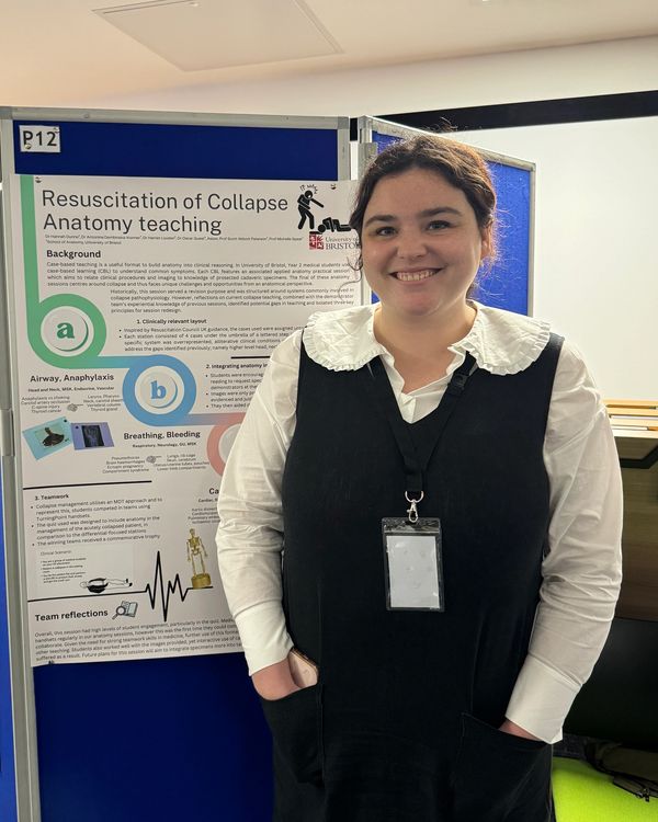
Hannah Dunne, Antonina Dembinska-Kenner, Harriet Louden, Oscar Guest
School of Anatomy, University Bristol,
Bristol, England, United Kingdom
Case-based teaching is a useful format to build anatomy into clinical reasoning. In University of Bristol, Year 2 medical students use case-based learning to understand common symptoms. This features an associated applied anatomy practical session which aims to relate clinical procedures and imaging to knowledge of prosected cadaveric specimens. The final anatomy session centres around collapse and thus faces unique challenges and opportunities from an anatomical perspective. As collapse causes span multiple organ systems, it affords a valuable opportunity for revision yet can be difficult to structure. Reflections on current collapse teaching were combined with experiential knowledge of previous sessions. This identified potential gaps in teaching and isolated key three principles for session redesign. 1. Clinically relevant layout; Inspired by Resuscitation Council UK guidance, the cases used were assigned unto A-E rotational stations. 2. Integrating anatomy into investigation; Students were encouraged to use the cadaveric specimens and pre-reading to request radiology/clinical images from the demonstrators at their station. Images were provided when anatomical reasoning was shown and then used to consider management options. 3. Teamwork; Collapse management utilises an MDT approach and to represent this, students competed in teams using TurningPoint handsets. The quiz used was designed to include anatomy in the management of the acutely collapsed patient, in comparison to the differential-focused stations. Reflections from this session highlighted a need for more use of specimens within this context and excellent student engagement with the competitive quiz. Future plans for this session will aim to integrate specimens into task-based activities.
Keywords: collapse, education, CBL
Elena Patera
Human Anatomy Resource Centre, University of Liverpool,
Liverpool, United Kingdom
Becoming an anatomy educator was a career option I never knew existed. My journey into higher education began when I moved to the United Kingdom in 2015 to pursue an undergraduate degree in Biological Sciences (Study Abroad) at Lancaster University. For my second year I moved to the United States where I had the opportunity to attend the Body Worlds Exhibition. Seeing dissected plastinated bodies for the first time ignited my love for anatomy and led me to pursue a Human Anatomy MSc at the University of Edinburgh. In 2020, I started working as an anatomy demonstrator at the University of Birmingham where I gained experience in small group teaching sessions, prosection-based practicals, and histology lectures. In 2021 I was awarded the Anatomy Medical Traineeship Scholarship by University College Dublin where I conducted a research master’s focusing on the use of multimedia resources for neuroanatomy education and worked part-time as a demonstrator facilitating dissection-based practicals. In June 2022, I started working as a Lead Demonstrator at St George’s University of London where I was responsible for delivering prosection-based practicals and lectures. While working at SGUL, I completed a postgraduate certificate in healthcare and biomedical education. In October 2023, I moved to Liverpool to embark on a long new journey as I started my PhD in Neuroanatomy while working part-time as a Doctoral Academic Teacher. Having studied and worked at different institutions allowed me to gain valuable personal and professional experiences, experience different cultures and individuals, and develop resilience and perseverance.
Keywords: anatomy, career reflection, academia
Reece Foster
University of Sunderland, School of Medicine,
Sunderland, United Kingdom
bi53qo@student.sunderland.ac.uk
Teaching is an essential part of any doctor’s role, but there is insufficient training given on how to teach and little to no opportunity for feedback on teaching skills. Since teaching is so important, why is it not implemented into the medical curriculum sufficiently to represent this? This poster investigates the benefits of near peer tutors (NPT) in neuro-anatomy laboratory sessions. The poster discusses 3 studies from different institutes implementation of NPT neuroanatomy teaching. Study 1 and 2 are quantitative. Study 1 shows very positive feedback from second year medical students of NPT when asked to rate their experiences. Study 2 statistics outline that early year’s medical students highly value their NPT experiences as it creates a positive learning community and enhances understanding. But as students’ progress through the year groups, there is less favorable responses to receiving NPT. This is evidence of pre-clinical years medical students being the most suitable recipients of near peer tutoring. Study 3 is qualitative interviews of those who have been near peer tutors, revealing professional development, in terms of interpersonal skills, teaching skills and academic skills. Linking all of this to future careers as doctors, having a key role in educating junior doctors, medical students, and other health professionals. Discussion and synthesis of date follows these studies. The conclusion is that preclinical years should be taught by NPT’s from clinical years in neuro anatomy laboratory sessions to enhance understanding and engagement of tutees, as well as enhancing the skills of the NPT’s themselves.
Keywords: education, near-peer, laboratory